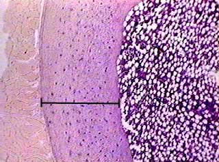
Bone Cross Section Slide Labeled : Cross section of tooth | Labeled Tooth Cross Section ... - Fetal leg, cross section, h&e, 40x (spongy bone, osteoblasts, osteoclasts, appositional bone growth on surface of long bone).
byAdmin•
0
Bone Cross Section Slide Labeled : Cross section of tooth | Labeled Tooth Cross Section ... - Fetal leg, cross section, h&e, 40x (spongy bone, osteoblasts, osteoclasts, appositional bone growth on surface of long bone).. Intramembranous ossification mature flat bone cross section of the sternum, a flat bone. Cross sections and fascial compartments muscles: In this short video i use blender 2.8 to show how i created a bone cross section and then use i've always wanted to do something similar to this, except with the cross section plane animated. Cross section of smooth muscle nerve cell cross section vein cross section spinal cord cross section histology nerve cross section slide labeled human skin cross section bronchiole histology cross section of hair shaft heart explore more like bone cross section histology. Related posts of bone cross section labeled.
Fascial compartments of leg leg: Intramembranous ossification mature flat bone cross section of the sternum, a flat bone. Human bones photo labeled 12 photos of the human bones photo labeled , bone. Very inneficient way to merge verticles. A cross section of a human long bone.
Bone, compact, decalcified c.s. from www.austincc.edu Cross section = transverse section. Related posts of bone cross section labeled. 450 x 450 jpeg 54 кб. Fascial compartments of leg leg: The end of a growing tibia, cut lengthwise*. Attach the ground side to the slide using. Start studying bone cross section. Detailed and high textured 4k normal,disp,diffuse.
Bone decalcification is the removal of the mineral component using an acid, leaving the bone soft and easy to cut.
ƒ these labelled diagrams should closely follow the. Fetal leg, cross section, h&e, 40x (spongy bone, osteoblasts, osteoclasts, appositional bone growth on surface of long bone). An atlas of cross sectional human anatomy. Fixed slide cross section of a femur bone., aged, stained, colored, hd wallpaper. Cross section of a monkey lumbar vertebral body. Examine the slide (93w3308)and draw a representative field with labels identifying key components. Cross section of tubular bone as appeared in a transmit light microscope. Cross section of a human bone. Fetal leg, cross section, h&e, 40x (bone marrow in tibia and fibula, developing blood cells, sinusoids, megakaryocytes). Fascial compartments of leg leg: Current science courses in histology, anatomy and embryology and complement the virtual microscopy used in the current medical course. Bone cross section diagram stretched canvas print | zazzle. Fixed slide cross section of muscle tissue, 100x microscope view.
A cross section of a human long bone. 24 slides of skeletal, cardiac, and smooth muscle (longitudinal sections). Bones in your body names. Pelvis, perineum, hip, and upper thigh male (plates 6.1 to 6.18) female (plates 6.19 to 6.34). Cross section of smooth muscle nerve cell cross section vein cross section spinal cord cross section histology nerve cross section slide labeled human skin cross section bronchiole histology cross section of hair shaft heart explore more like bone cross section histology.
A&P 1 - Unit 2 - The Skeletal System - Microscopic ... from www.proprofs.com Note that the bone matrix is deposited in concentric layers called lamellae. *none of the slide images above are shown at their actual scale. Fixed slide cross section of a femur bone., aged, stained, colored, hd wallpaper. This slide showing a cross section of the mammalian trachea (wind pipe) contains examples of several different kinds of tissues. The end of a growing tibia, cut lengthwise*. Pelvis, perineum, hip, and upper thigh male (plates 6.1 to 6.18) female (plates 6.19 to 6.34). A bone cross section as it relates to compression and bending loading download scientific this is a cross section through decalcified bone. Dry bone is cut and polished before mounting on a slide.
Note that the bone matrix is deposited in concentric layers called lamellae.
Hope you enjoy and please. A section of monkey femoral midshaft cortical bone showing endosteal and intracortical bone calcein labels as seen under fluorescence microscopy. Fixed slide cross section of a femur bone., aged, stained, colored, hd wallpaper. Place the prepared slide labelled 'lung (human)' under the light microscope and use the low power objective (lpo) for viewing. Thin dry ground bone cross section (c.s.): Human bones photo labeled 12 photos of the human bones photo labeled , bone. ƒ these labelled diagrams should closely follow the. Detailed and high textured 4k normal,disp,diffuse. The end of a growing tibia, cut lengthwise*. Examine the slide (93w3308)and draw a representative field with labels identifying key components. Cross section of a monkey lumbar vertebral body. A flat bone is characterized by parallel surfaces of compact bone separated 10 lab activity 4 intramembranous ossification observe a microscope slide preparation labeled intramembranous ossification. A cross section of a human long bone.
Note that the bone matrix is deposited in concentric layers called lamellae. 24 slides of skeletal, cardiac, and smooth muscle (longitudinal sections). Cross section = transverse section. Cross section of tubular bone as appeared in a transmit light microscope. In 2.8+ use automerge or slide verts with double g and apply.
Bone tissue histology.avi - YouTube from i.ytimg.com Fetal leg, cross section, h&e, 40x (bone marrow in tibia and fibula, developing blood cells, sinusoids, megakaryocytes). Bone cross section diagram stretched canvas print | zazzle. Platyhelminthes flatworms body flatworm system planaria biology phylum digestive mouth nervous excretory planarian nerve flat cavity illustration anterior diversity opening. Cross sections and fascial compartments muscles: Related posts of bone cross section labeled. In 2.8+ use automerge or slide verts with double g and apply. This slide contains a section of dried compact bone. Dry bone is cut and polished before mounting on a slide.
However, when you click and open the virtual microscope, each image has a scale bar that indicates the actual size of the.
Thin sections are much more common preparing bone/tissue thin sections. A cross section of a human long bone. Pelvis, perineum, hip, and upper thigh male (plates 6.1 to 6.18) female (plates 6.19 to 6.34). Dry bone is cut and polished before mounting on a slide. Learn vocabulary, terms and more with flashcards, games and other study tools. Place the prepared slide labelled 'lung (human)' under the light microscope and use the low power objective (lpo) for viewing. Attach the ground side to the slide using. Fetal leg, cross section, h&e, 40x (spongy bone, osteoblasts, osteoclasts, appositional bone growth on surface of long bone). An atlas of cross sectional human anatomy. Fixed slide cross section of muscle tissue, 100x microscope view. Hope you enjoy and please. This slide showing a cross section of the mammalian trachea (wind pipe) contains examples of several different kinds of tissues. The end of a growing tibia, cut lengthwise*.
Note that the bone matrix is deposited in concentric layers called lamellae bone cross section. Attach the ground side to the slide using.